info@kingyork.net
+44 7478 549592
info@kingyork.net
+44 7478 549592
Magnetic resonance imaging, commonly known as MRI, is a medical imaging technique that utilizes strong magnetic fields and radio waves to generate detailed images of the organs and tissues within the body. This non-invasive procedure is widely used for diagnosing various medical conditions, providing critical insights into the structure and function of internal systems.
Magnetic resonance imaging is a non-invasive diagnostic method.
This imaging technique utilizes magnetic fields and radio waves to generate detailed images of internal body structures.
It is widely employed in medical settings for its ability to visualize soft tissues without the need for surgical intervention.
MRI is preferred for its safety and lack of ionizing radiation.
MRI is particularly valuable in the fields of neurology, orthopedics, and oncology, as it offers high-resolution images without the use of ionizing radiation. The technology allows healthcare professionals to assess abnormalities, monitor treatment progress, and plan surgical interventions with greater accuracy.
Magnetic resonance imaging (MRI) is a diagnostic technique that utilizes magnetic fields and radio waves to generate detailed images of the body's internal structures.
One of the significant advantages of MRI is that it does not involve the use of ionizing radiation, making it a safer alternative for patients, particularly for those requiring multiple imaging sessions.
This non-invasive imaging modality is particularly valuable in assessing soft tissues, providing critical information for the diagnosis and management of various medical conditions.

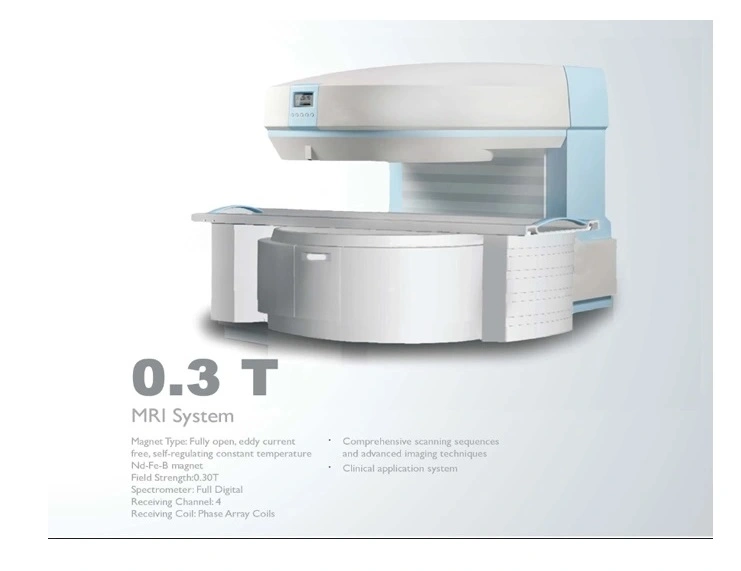
Magnetic resonance imaging, commonly referred to as MRI, is a sophisticated medical imaging technique that utilizes a magnetic field of 0.3 Tesla to produce detailed images of the internal structures of the body. This specific strength of the magnetic field allows for the visualization of soft tissues, making it particularly useful in diagnosing various medical conditions. The 0.3T MRI system is often employed in clinical settings where high-resolution images are necessary for accurate assessment and treatment planning, providing healthcare professionals with critical information about the patient's anatomy.
The application of a 0.3 Tesla magnetic field in MRI technology enhances the contrast of images, enabling the detection of abnormalities that may not be visible with lower field strengths. This imaging modality is non-invasive and does not involve ionizing radiation, making it a safer alternative for patients requiring frequent imaging. As a result, MRI has become an essential tool in modern medicine, facilitating the diagnosis of conditions such as tumors, neurological disorders, and musculoskeletal injuries, while also guiding therapeutic interventions.

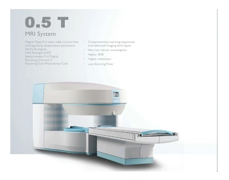
Magnetic resonance imaging, commonly referred to as MRI, is a sophisticated medical imaging technique that utilizes a magnetic field of 0.5 Tesla to produce detailed images of the internal structures of the body. This specific strength of the magnetic field allows for the visualization of soft tissues, making it particularly useful in diagnosing various medical conditions. The 0.5T MRI system is often employed in clinical settings where high-resolution imaging is required, yet the lower magnetic field strength can be advantageous for certain patient populations.
The application of a 0.5T MRI system is significant in various medical fields, including neurology, orthopedics, and oncology. By generating high-quality images, this imaging modality aids healthcare professionals in making accurate diagnoses and developing effective treatment plans. Furthermore, the use of a lower magnetic field can enhance patient comfort and safety, as it reduces the risk of adverse reactions associated with higher field strengths. Overall, the 0.5T MRI represents a valuable tool in modern medicine, contributing to improved patient outcomes through precise imaging capabilities.

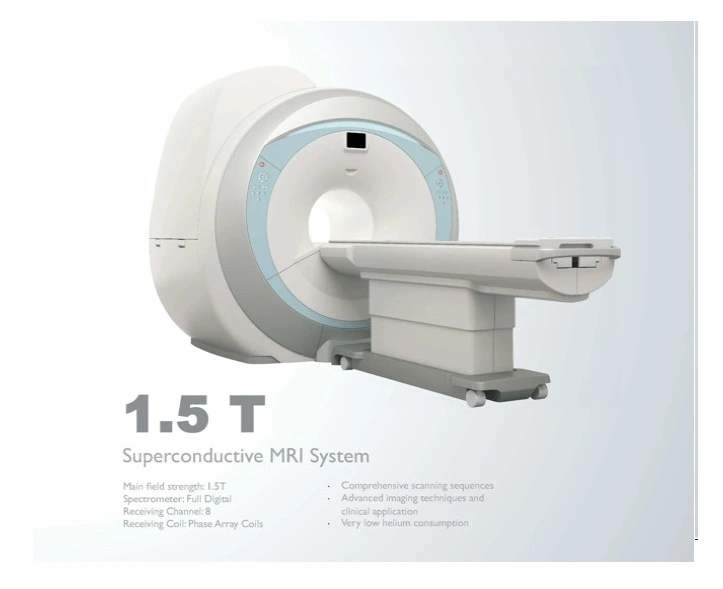
Magnetic resonance imaging, commonly referred to as MRI, operates at a strength of 1.5 Tesla. This advanced imaging technique utilizes powerful magnetic fields and radio waves to generate detailed images of the internal structures of the body. The 1.5T strength is particularly effective for a wide range of diagnostic applications, providing high-resolution images that assist healthcare professionals in identifying various medical conditions.
The 1.5 Tesla MRI system is widely used in clinical settings due to its balance of image quality and patient safety. It is capable of producing clear images of soft tissues, organs, and even the brain, making it an invaluable tool in modern medicine. The technology has evolved significantly, allowing for faster scan times and improved patient comfort, which enhances the overall experience for individuals undergoing this essential diagnostic procedure.
This imaging technique is particularly effective for visualizing soft tissues, making it invaluable for diagnosing conditions related to the brain, spinal cord, and joints.